Specialist in Oral and Maxillofacial Surgery
Plastic Surgery of the Face
Specialist Dentist for Oral Surgery
Head of the Department for Oral, Maxillofacial
and Plastic Facial Surgery
Helios Hospital Berlin Buch
Expert witness for the Dental Chambers of Berlin and Brandenburg
and the Medical Chamber of Brandenburg for Implantology and OMF Surgery
Authorised training supervisor for the Specialist Dentist
in Oral Surgery programme (3 years)
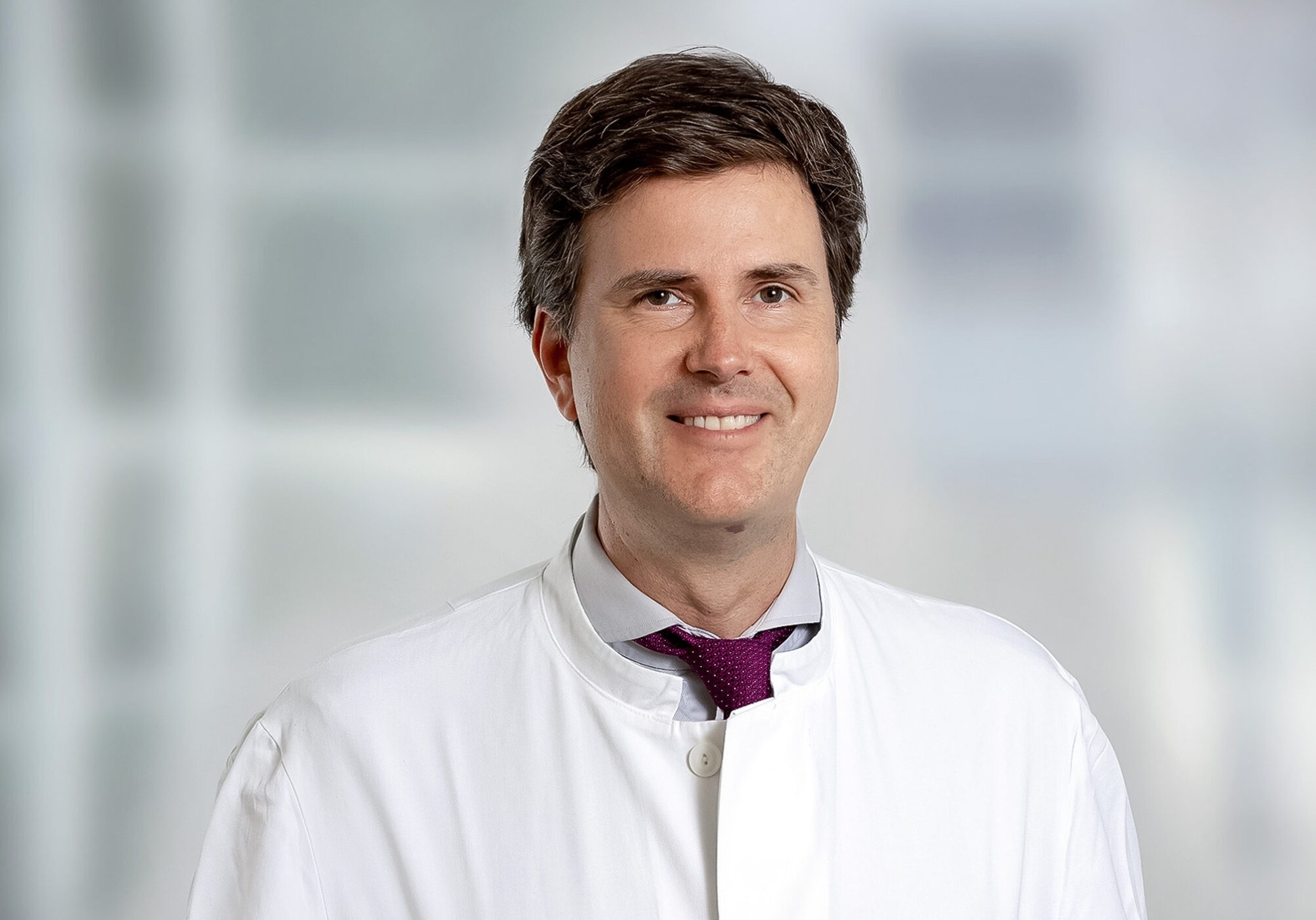
My name is Branko Šiniković, I am an oral and maxillofacial surgeon and oral surgeon. I cover the full spectrum of my specialties and practice both in hospital and on an outpatient basis.
I work as Head of the Department for OMF and Plastic Facial Surgery at Helios Hospital Berlin Buch and in my private practice in Berlin Kreuzberg.
I hope this website is interesting and helpful for you.
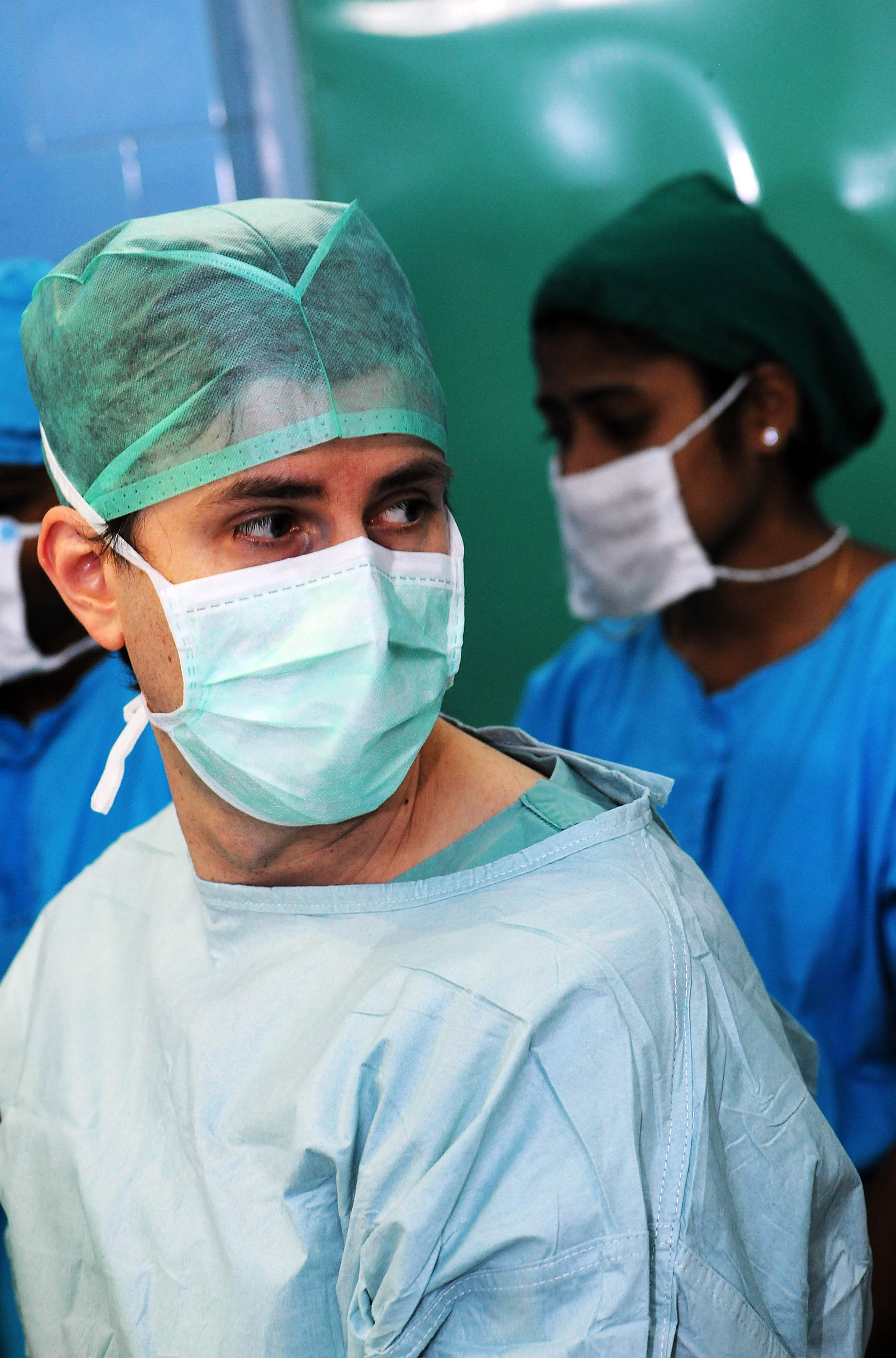
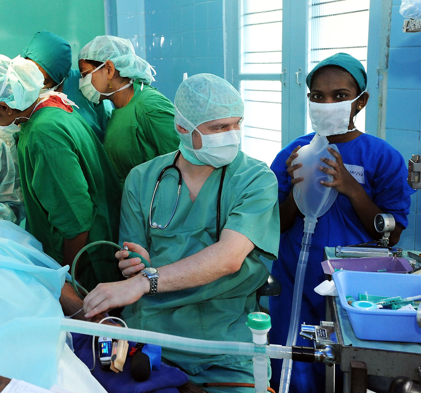
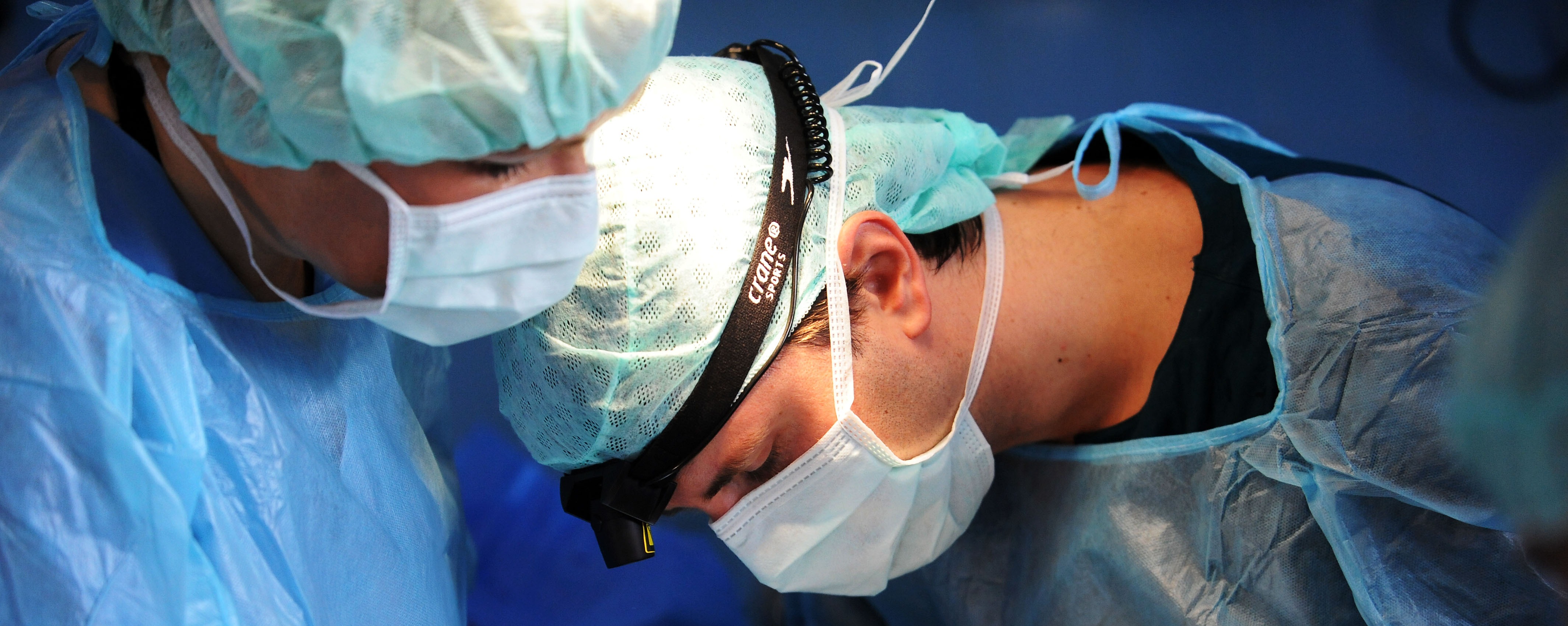
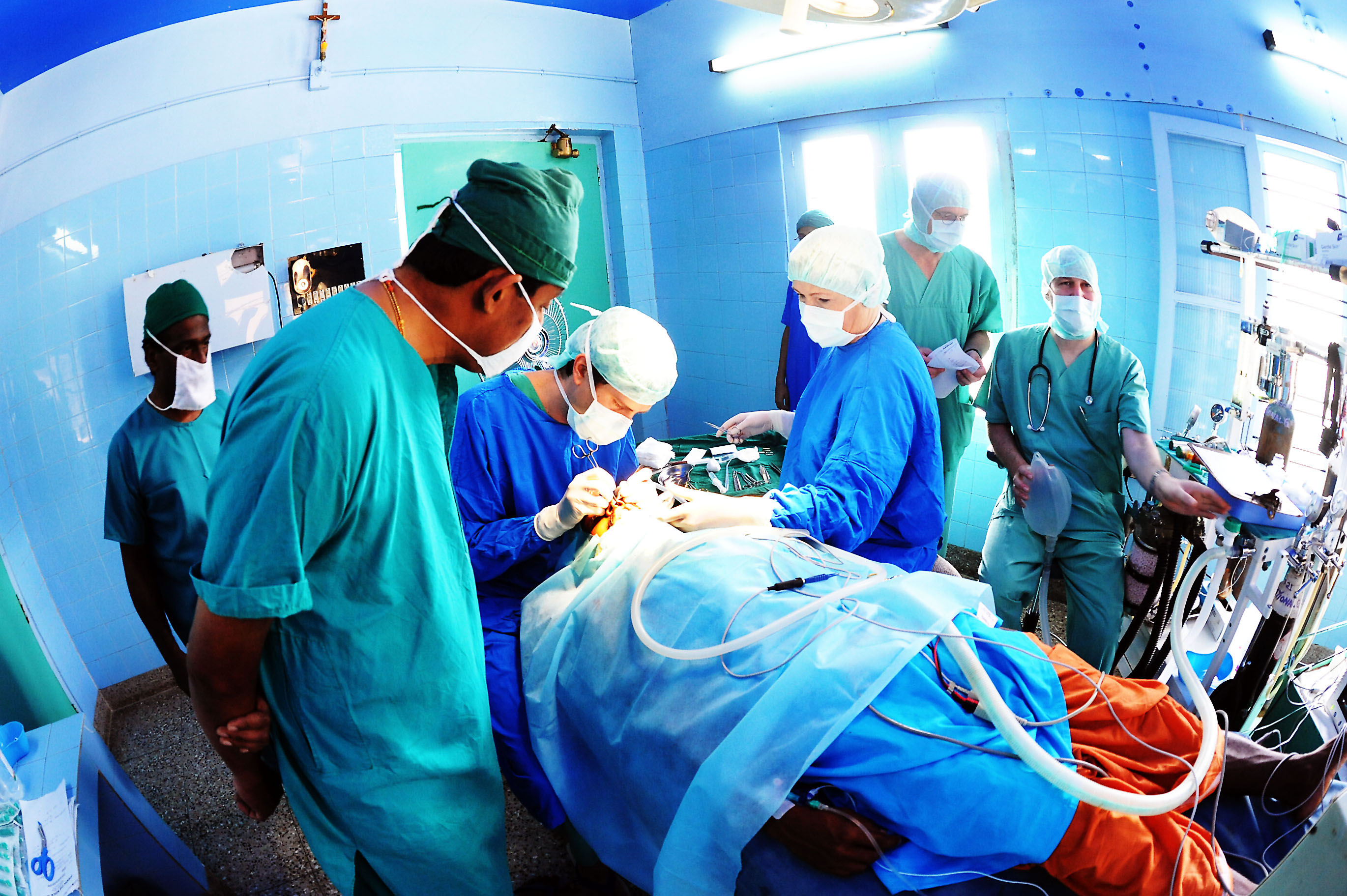
Charitable mission Adimali with Interplast, India. With kind permission of Oskar Neubauer (all rights reserved)
Orthognathic surgery is a specific subspecialty of oral and maxillofacial surgery, as no other medical discipline covers this area.
It is indicated when misalignments of the upper and/or lower jaw are present that cannot be adequately corrected by orthodontic treatment (braces) alone. The cause usually lies in a congenital malposition of the jaw bases relative to each other or to the skull base. This can only be influenced during growth, and even then only to a limited extent through orthodontic treatment.
Once growth is complete – roughly by the end of the teenage years – or in particularly severe cases of jaw misalignment, the tooth-bearing jaw segments are surgically repositioned to achieve functional and aesthetic harmony between the teeth of both jaws, the jaw bases, the temporomandibular joints and the rest of the face. This represents both a functional and an aesthetic improvement aimed at eliminating long-term wear of the masticatory system and its consequences. For this reason, when certain criteria are met, almost the entire therapy is covered by statutory health insurance.
For surgical planning I use digital systems to simulate the required jaw movements before the operation. This three-dimensional information is transferred into a plastic splint – a horseshoe-shaped acrylic block containing impressions of the teeth of both jaws. It helps locate the target position of the respective jaw during surgery and, at the same time, provides the patient with stable support afterwards, since the precise position of each individual tooth is only achieved during post-operative orthodontic treatment.
During the first eight weeks after surgery the jaw must consolidate and stabilise in its new position, so only soft food is permitted and chewing of solid food is not allowed. These jaw repositioning operations are performed under general anaesthesia at the hospital during a short inpatient stay.
Also known as genioplasty, this procedure aims to harmonise the facial profile and chin projection by repositioning and/or reshaping the bony chin in the middle and lower mandibular region. Genioplasty is usually performed only in the anterior region, between the exit points of the sensory nerves (interforaminally) and below the tooth roots, but in certain indications it can be extended to the mandibular angle (“chin-wing” after Albino Triaca).
Unlike repositioning of the entire lower jaw – which also changes the chin position – genioplasty modifies the chin region in isolation. It can be performed as a complement to mandibular repositioning when a more pronounced change in chin position is desired.
This term refers generally to surgical procedures involving the skin. In most cases, skin changes are treated surgically – for example, removal of benign and malignant skin tumours, moles, intra- or subcutaneous cysts, treatment of existing scars, and correction of disturbing skin folds or sagging tissue (e.g. eyelid lift – blepharoplasty).
Patients are typically referred to an oral and maxillofacial surgeon for dermatosurgical procedures when “sensitive” facial areas are involved – the eyelids, the outer ear, changes to the forehead and nasal skin, and proximity to the nostrils and lips. Even mere proximity to these areas can have a negative effect through later scar contraction, for instance causing a displaced eyelid (blepharoptosis). Likewise, removal of deeper skin changes can compromise the facial nerve and thus lead to paralysis of the mimetic musculature, which can be avoided through specialised surgical techniques.
These treatments take place in trusted, collegial collaboration between the treating dermatologist and the OMF surgeon.
Also referred to as oral surgery or dental surgery. It encompasses operations within the oral cavity involving teeth, tooth roots, as well as cystic and other pathological changes or bony overgrowths of the jaw bones and palate. Oral surgical procedures also include areas of the maxillary sinuses and the oral mucosa. These are very commonly performed outpatient operations, such as wisdom tooth removal, root tip resection and tooth extraction.
This field also includes dental implantology. As a surgical-prosthetic measure enabling the anchoring of fixed dental prostheses in the jaw bone, this subspecialty covers various measures for the reconstruction of defects of the jaw bone and mucosa, as well as the placement of dental implants that functionally integrate with the jaw bone as “artificial tooth roots” (osseointegration). These treatments are carried out in collegial collaboration and coordination with the subsequent prosthodontist.
Combined with one year of inpatient hospital training, I hold the full authorisation of the Berlin Dental Chamber for the three-year Specialist Dentist in Oral Surgery training programme.
A modern imaging technique from the field of dental radiology, used primarily in OMF surgery and dentistry. More recently, it is also gaining ground in orthopaedics and trauma surgery. It is a CT-like procedure but, since it is designed for hard tissue imaging (bones, teeth and cavities), it delivers a lower radiation dose than conventional computed tomography (CT).
Thanks to its high resolution, it is used for diagnostic purposes as well as – with appropriate software – for the simulation and planning of surgical procedures, such as implant placement or jaw repositioning. The three-dimensional CBCT data set is combined with other digital 3D data sets, such as STL files, achieving high precision of the computed models. At the end of the planning process, the 3D planning data is transferred to the surgical site as drilling guides or splints using a 3D printer.
This is a sleep disorder in which breathing repeatedly stops during sleep, significantly impairing oxygen supply to the brain and causing other negative health effects.
After the diagnosis is confirmed in a suitable sleep laboratory by determining the AHI (an index describing the number of breathing pauses), and ideally after identifying the relevant airway obstruction, treatment is initiated. The gold standard is keeping the airway open using a positive-pressure ventilation mask (CPAP) worn over the nose and mouth during sleep. Some patients tolerate this therapy poorly, as small leaks at the mask edge can cause air to escape and wake the patient. Some patients find wearing a mask pressed onto the face under pressure very disturbing.
If the relevant obstruction is located at the level of the base of the tongue or the mid-pharynx, a mandibular advancement device (MAD) – which creates more space behind the tongue by advancing the lower jaw – can provide relief. Such an OSAS splint (also called a “snoring splint”) must be individually fabricated, as it is tooth-borne. This applies both to the fit on the teeth and to the individually determined advancement that is tolerated by the teeth and temporomandibular joints. After fitting, regular follow-up checks and adjustments should be carried out.
If the cause of the airway narrowing is a jaw misalignment (dysgnathia) – typically a retrusion – this can be very effectively corrected through orthognathic surgery. As a side benefit, the dental and jaw misalignment would also be corrected. When such circumstances are the cause of nocturnal breathing pauses, jaw repositioning represents the most effective surgical measure (after tracheotomy) for their elimination. A severe OSAS can therefore be considered a further indication for jaw repositioning surgery.
Here you can download specific instructions and recommendations that are useful before or after surgery. You will receive the corresponding access code at the practice.
As an expert witness in the field of oral, maxillofacial and plastic facial surgery, oral surgery and dental implantology, I regularly deal with medico-legal questions.
Expert reports can be commissioned privately or requested through solicitors or courts. It is important that, before commissioning a report, a clear idea exists of the purpose for which the report is to be prepared and used. Equally important is to formulate clear questions to be addressed, in order to keep workload and associated costs low. Please note that expert reports are prepared outside of treatment hours, i.e. in my “free time.” Depending on the availability of relevant documents, the effort involved and the number of questions to be addressed, preparation of a report may take several months.
In the event of potential legal disputes, please note that an expert report commissioned by one party is regarded by the court as a submission of that party, unlike a court-appointed expert opinion, which follows a specific procedure and requires the consent of all parties.
Expert activity in a case precludes subsequent therapeutic activity in the same case due to potential conflicts of interest. The same applies in reverse.
Please print the appropriate form and give it to the patient, filled out.
Print referral and hospital admission formPlease choose a date and time for a consultation. The consultation takes place at:
Bergmannpraxis, Bergmannstraße 102, D-10961 Berlin Kreuzberg.
If you cannot attend in person, please choose Send email. Files can be provided via KIM, email, or Doctolib.
Online AppointmentsWe aim to respond to your email within 3–4 working days.
This website has an SSL certificate.